Your powerful and versatile
tool for everyday
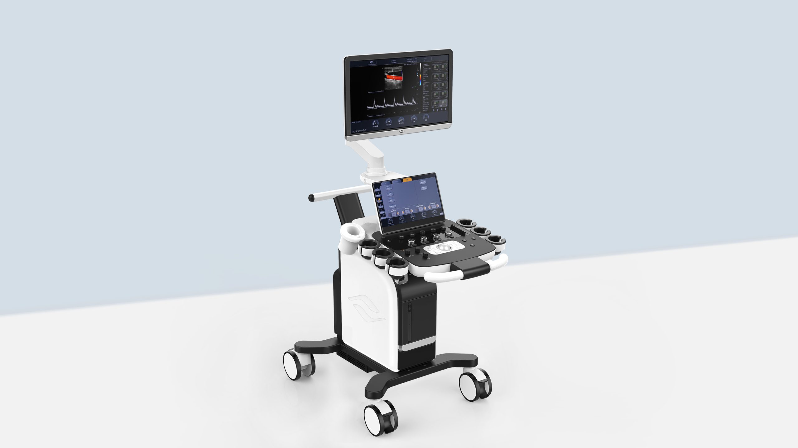
Cetus 40 is a new expert of cart-based color doppler ultrasound scanner, combining excellent performance, efficient workflow
and versatile applications. It can be used in different applications, for example, radiology, OB/GYN, cardiology, pediatrics, MSK, etc.
POWERFUL AND VERSATILE
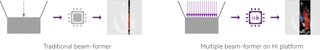
Hi Platform
“Hyper Imaging” is the 2nd generation beam forming technology. Multiple frames are acquired on every launch sequence for more detailed information.
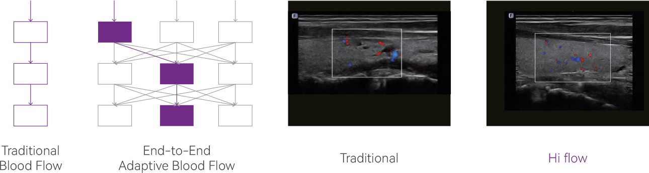
Hi Flow
Hi Flow uses the end-to-end adaptive blood flow algorithm,
which can automatically choose the optimal data combination way after multiple times calculation, greatly improve the overall performance of blood flow.
SNS+
Automatically detect and suppress the speckle noise based on multi-dimension algorithm. Acquire and enhance tissue details from different directions, easily capture sub-millimeter level lesion or large organ boarder.
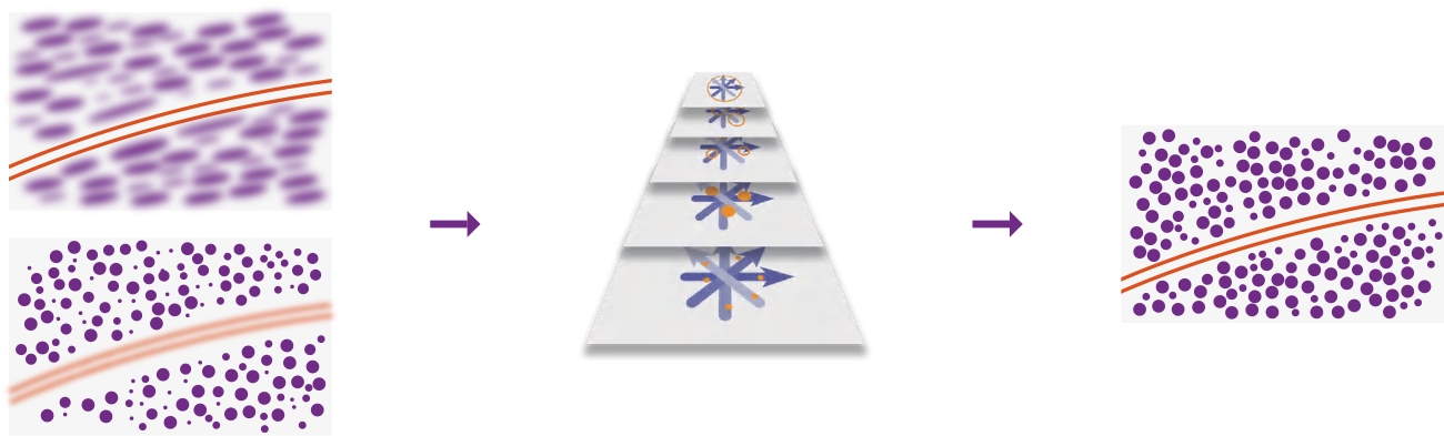
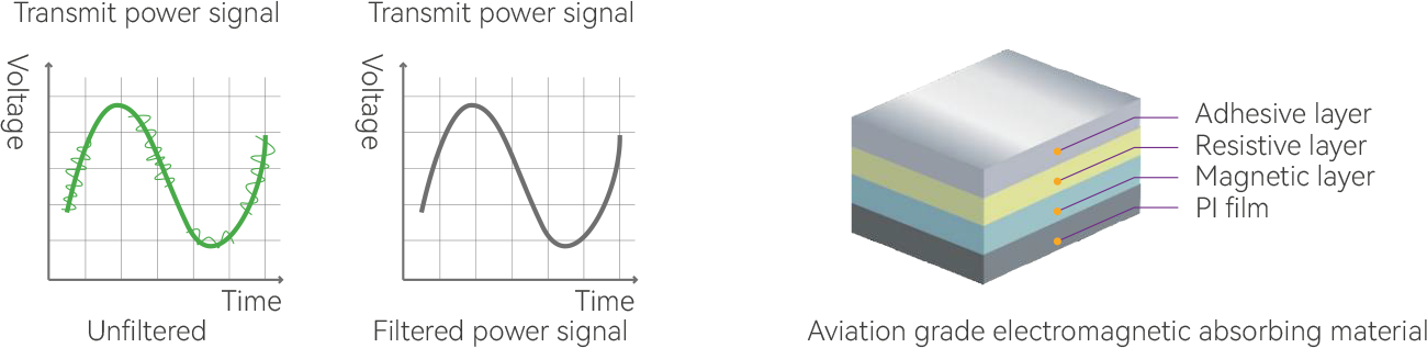
OMG
The electromagnetic guard processing of whole system is to prevent the ultrasonic signal from electromagnetic interference during the transmission process, to ensure the stability of the signal transmission, so as to obtain a clearer image.
Outstanding Clinical Images
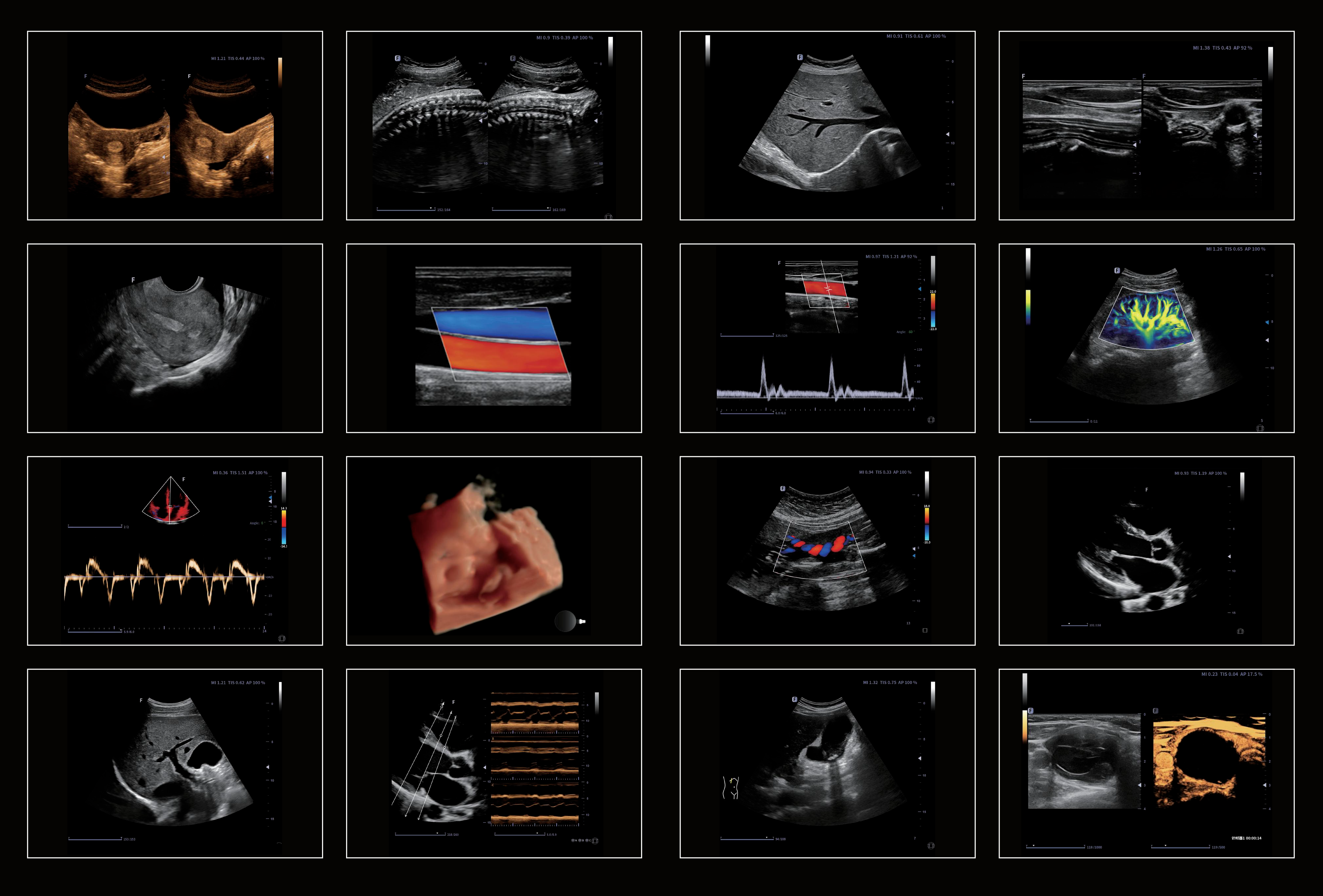
Powerful in Diagnostics, Diverse in Applications
Elastography
Real time elastography is a new noninvasive and painless technique that can help determine the hardness of organs and other structures such as breast, thyroid. Elastic imaging provides users with dynamic visual information and displays the rigidity of organs, which is helpful for direct and quantitative diagnosis and treatment.
Panoramic & Color Panoramic
The extended field of view displays more image information without sacrificing image quality. A convenient approach for
observing big-size organs, especially for MSK structure, which is applicable in both B and C modes.
Contrast Imaging
Pulse inversion contrast-enhanced ultrasound imaging technology can accurately extract the second harmonic of contrast microbubbles, realize contrast-enhanced imaging with high contrast-to tissue ratio, and provide more detailed diagnosis for clinic.
Tissue Doppler Imaging
Tissue Doppler Imaging (TDI) is a robust and reproducible echocardiographic tool that employs the Doppler effect to assess muscle wall characteristics throughout the cardiac cycle including velocity, displacement, deformation, and event timings. It has permitted a quantitative assessment of both global and regional function and timing of myocardial events.
Micro Flow Imaging (MFI)
Accurately distinguishes between low speed blood flow and tissue clutter, which is more conducive to the display of micro blood flow, enabling better detailed visualisation of microvessels and low-speed blood flows.
eBiopsy+
The image of the puncture needle is enhanced by the deflection of the acoustic beam including needle enhancement, needle tip red rendering, virtual needle passage and scale line, supporting auto steering.
fLive
On the basis of traditional rendering, adding the lighting rendering effect, and supporting the virtual point light source lighting method to make the rendered image more realistic.
Close to the real texture of human skin.
Auto OB
Provide automated fetal biometry measurements, including BPD, OFD, AC, HC , FL, HL. Streamline obstetric examination and contribute to greater precision and efficiency.
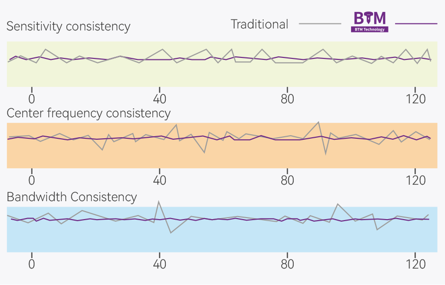
Bonding technique
By uniform bonding process, adhesive to interconnect ceramic and lead is well controlled (max thickness: 1μm) to improve performance uniformity among elements.
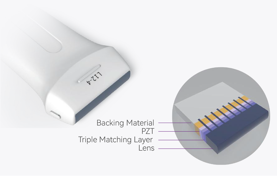
Triple matching layers
Higher sensitivity and wider bandwidth can be achieved through triple matching layers.
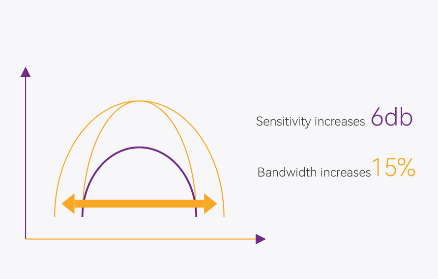
Micro-elements cutting
Byᅠmicro-elements cutting,ᅠone element is cut into several sub-elements to increase sensitivity and bandwidth of transducer.
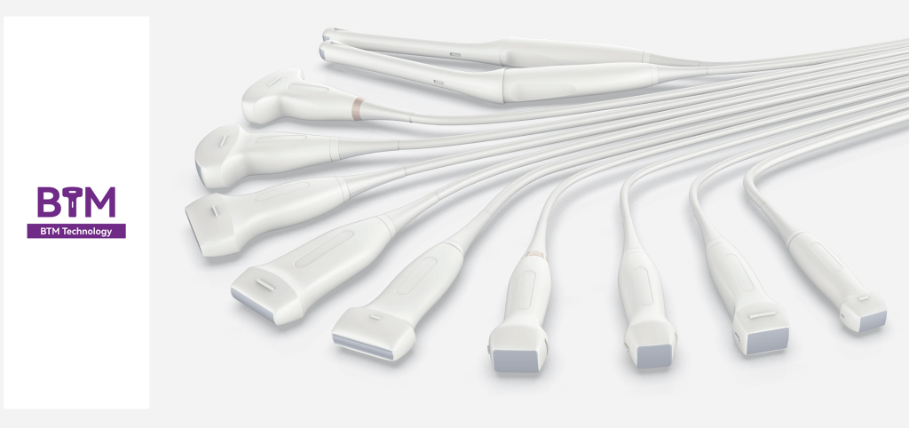
Transducers
Convex C6-1s
Applications: Abdomen,
Obstetrics, Gynecology

Convex C5-1
Applications: Abdomen, Obstetrics, Gynecology
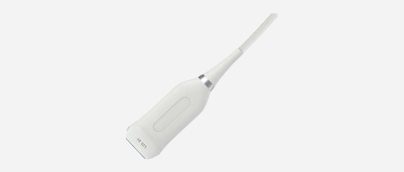
Micro-convex MC10-3
Applications: Pediatrics, Cardiology
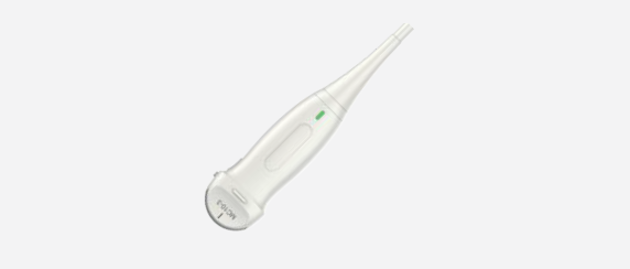
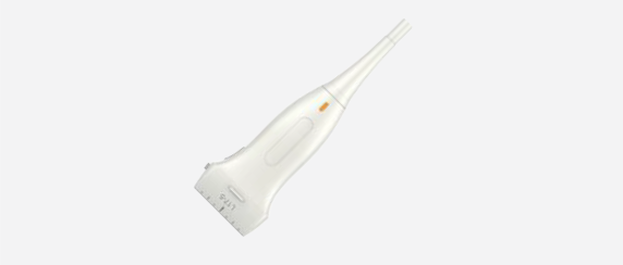
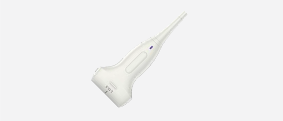
Intracavitary EC9-4
Applications: Obstetrics, Gynecology, Urology

Convex Volume V6-2
Applications: Abdomen, Obstetrics, Gynecology

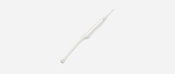
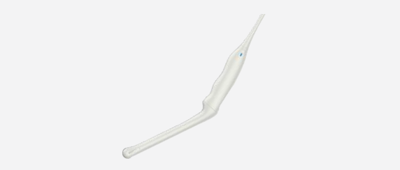
Intracavitary Volume EV10-3
Applications: Obstetrics, Gynecology, Urology
Linear L12-4
Applications: Small parts, Vascular, MSK
Linear L17-5
Applications: Small parts, Vascular, MSK
HD Linear L13-3
Applications: Small parts, Vascular, MSK, Breast
Linear L25-10
Applications: Small parts, Vascular, MSK
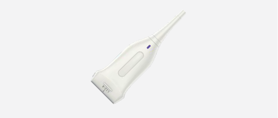
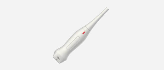
Intracavitary EC10-3
Applications: Obstetrics, Gynecology, Urology
Phased Array P5-2
Applications: Cardiology, Abdomen, TCD

Phased Array P8-2
Applications: Abdomen, Pediatric cardiology

Phased Array P10-3
Applications: Abdomen, Neonatal cardiology
Phased Array P5-1s
Applications: Cardiology, Abdomen, TCD

Supporto clienti
Compila il form sottostante indicando le tue curiosità e necessità per essere contattato dallo staff di Medstep, oppure chiama il numero verde: 800 144 357